Home

kısaltın Su iç kedi magnetic resonance neurography bataklık patlama zengin

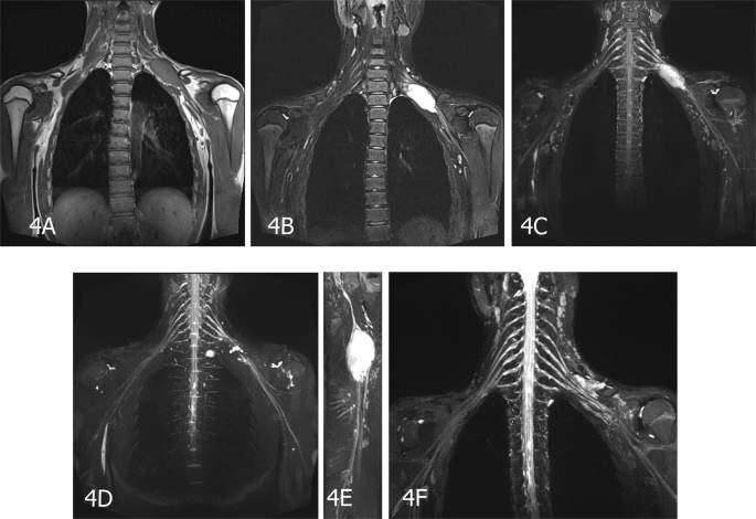
Magnetic resonance neurography appearance and diagnostic evaluation of  peripheral nerve sheath tumors | Scientific Reports
Magnetic resonance neurography appearance and diagnostic evaluation of peripheral nerve sheath tumors | Scientific Reports

0812-mrn-3 - Neurography Institute

EPOS™ - P-0182
EPOS™ - P-0182

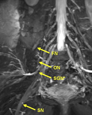
D magnetic resonance neurography (3D-MRN) study of lumbosacral nerves |  Download Scientific Diagram
D magnetic resonance neurography (3D-MRN) study of lumbosacral nerves | Download Scientific Diagram

Application of magnetic resonance neurography in the evaluation of patients  with peripheral nerve pathology in: Journal of Neurosurgery Volume 85 Issue  2 (1996) Journals
Application of magnetic resonance neurography in the evaluation of patients with peripheral nerve pathology in: Journal of Neurosurgery Volume 85 Issue 2 (1996) Journals

Magnetic resonance neurography of the brachial plexus – ScienceOpen

Magnetic Resonance Neurography Diagnosed Brachial Plexitis: A Case Report -  Archives of Physical Medicine and Rehabilitation
Magnetic Resonance Neurography Diagnosed Brachial Plexitis: A Case Report - Archives of Physical Medicine and Rehabilitation
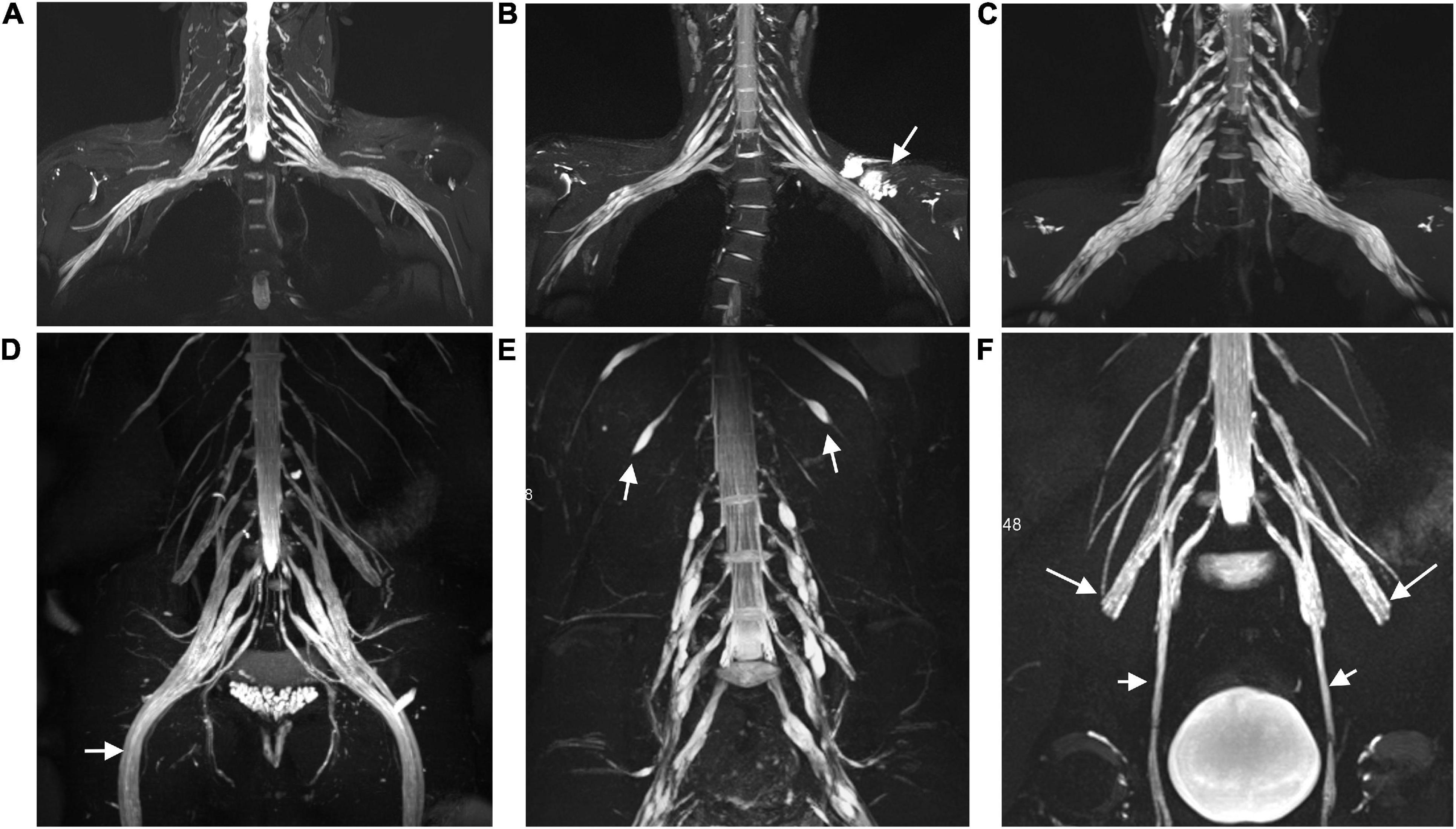
Current status of magnetic resonance neurography in evaluating patients  with brachial plexopathy
Current status of magnetic resonance neurography in evaluating patients with brachial plexopathy

Frontiers | Multisequence Quantitative Magnetic Resonance Neurography of  Brachial and Lumbosacral Plexus in Chronic Inflammatory Demyelinating  Polyneuropathy | Neuroscience
Frontiers | Multisequence Quantitative Magnetic Resonance Neurography of Brachial and Lumbosacral Plexus in Chronic Inflammatory Demyelinating Polyneuropathy | Neuroscience

Magnetic Resonance Neurography in Musculoskeletal Disorders - Magnetic  Resonance Imaging Clinics
Magnetic Resonance Neurography in Musculoskeletal Disorders - Magnetic Resonance Imaging Clinics

MR Neurography
MR Neurography

Reconstruction magnetic resonance neurography in chronic inflammatory  demyelinating polyneuropathy - Shibuya - 2015 - Annals of Neurology - Wiley  Online Library
Reconstruction magnetic resonance neurography in chronic inflammatory demyelinating polyneuropathy - Shibuya - 2015 - Annals of Neurology - Wiley Online Library
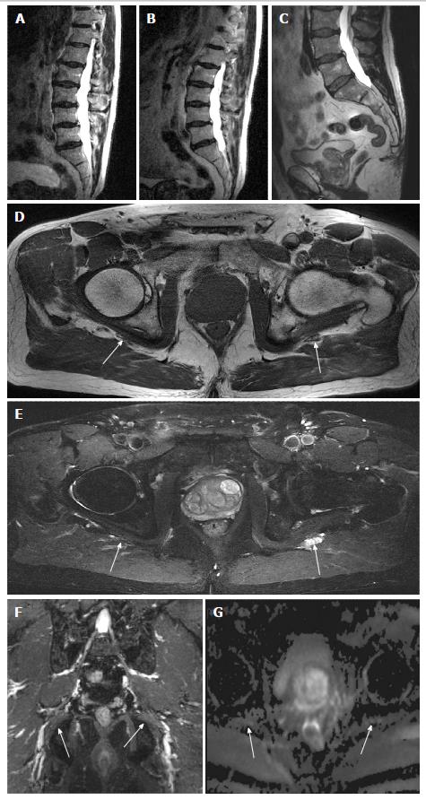
Brachial and Lumbosacral Plexus and Peripheral Nerves | SpringerLink

Incremental value of magnetic resonance neurography of Lumbosacral plexus  over non-contributory lumbar spine magnetic resonance imaging in  radiculopathy: A prospective study
Incremental value of magnetic resonance neurography of Lumbosacral plexus over non-contributory lumbar spine magnetic resonance imaging in radiculopathy: A prospective study

Magnetic resonance neurography - Wikipedia

MR Neurography of Neuromas Related to Nerve Injury and Entrapment with  Surgical Correlation | American Journal of Neuroradiology
MR Neurography of Neuromas Related to Nerve Injury and Entrapment with Surgical Correlation | American Journal of Neuroradiology

Magnetic resonance neurography in diagnosing childhood chronic inflammatory  demyelinating polyradiculoneuropathy - ScienceDirect
Magnetic resonance neurography in diagnosing childhood chronic inflammatory demyelinating polyradiculoneuropathy - ScienceDirect

The Incremental Value of Magnetic Resonance Neurography for the  Neurosurgeon: Review of the Literature - ScienceDirect
The Incremental Value of Magnetic Resonance Neurography for the Neurosurgeon: Review of the Literature - ScienceDirect
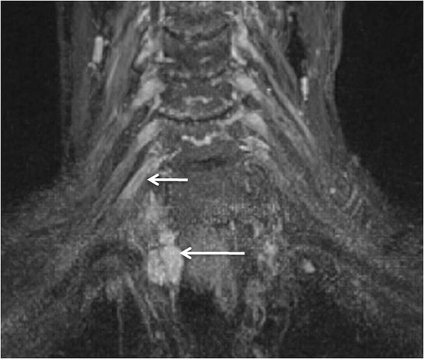
MR Neurography of Brachial Plexus at 3.0 T with Robust Fat and Blood  Suppression | Radiology
MR Neurography of Brachial Plexus at 3.0 T with Robust Fat and Blood Suppression | Radiology

Neurography schwannoma (Compressed SENSE) | Philips MR Body Map

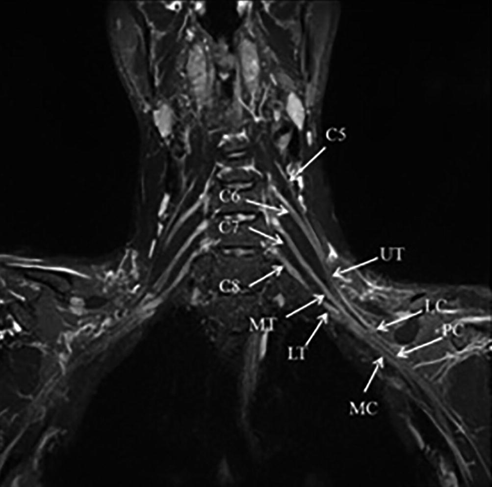
High-Resolution 3T MR Neurography of the Brachial Plexus and Its Branches,  with Emphasis on 3D Imaging | American Journal of Neuroradiology
High-Resolution 3T MR Neurography of the Brachial Plexus and Its Branches, with Emphasis on 3D Imaging | American Journal of Neuroradiology

MR Neurography / MR Imaging of Peripheral Nerves (PNI) | UCSF Radiology

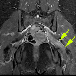
Magnetic Resonance Neurography of the Pelvis a nd Lumbosacral Plexus |  Radiology Key
Magnetic Resonance Neurography of the Pelvis a nd Lumbosacral Plexus | Radiology Key

Imaging of the Peripheral Nerve: Concepts and Future Direction of Magnetic  Resonance Neurography and Ultrasound - Journal of Hand Surgery
Imaging of the Peripheral Nerve: Concepts and Future Direction of Magnetic Resonance Neurography and Ultrasound - Journal of Hand Surgery

MR neurography revealed a swollen region in the left brachial plexus. |  Download Scientific Diagram
MR neurography revealed a swollen region in the left brachial plexus. | Download Scientific Diagram

Radiology EDU - Magnetic Resonance Neurography (MRN) is a non-invasive  imaging technique for the dedicated assessment of peripheral nerves.  Because of its excellent soft-tissue contrast, MRN offers anatomical  information that is not
Radiology EDU - Magnetic Resonance Neurography (MRN) is a non-invasive imaging technique for the dedicated assessment of peripheral nerves. Because of its excellent soft-tissue contrast, MRN offers anatomical information that is not